Tideline Margate is quickly becoming one of the most talked-about coastal destinations for modern living, offering a unique blend of luxury, comfort, and natural beauty. Located in the vibrant city of Margate, this development stands out for its contemporary design and proximity to stunning waterfront views. Whether you are considering relocation, investment, or simply exploring new living options, Tideline Margate offers something for everyone.
In recent years, the demand for well-designed residential communities has increased significantly, and Tideline Margate meets these expectations with its thoughtful planning and premium amenities. This guide explores everything you need to know about Tideline Margate, from its location advantages to lifestyle benefits and investment potential.
What Is Tideline Margate?
Tideline Margate is a modern residential development designed to provide a comfortable and stylish coastal living experience. Known for its sleek architecture and community-focused layout, it caters to individuals and families seeking a balance between urban convenience and natural tranquility. The development emphasizes open spaces, scenic surroundings, and high-quality construction standards.
One of the standout features of Tideline Margate is its strategic location within Broward County. This positioning allows residents to enjoy easy access to nearby cities, beaches, shopping centers, and recreational facilities. The project has gained attention for offering a peaceful environment while still being connected to essential urban amenities.
Prime Location and Accessibility
The location of Tideline Margate plays a significant role in its growing popularity. Situated in Margate, residents benefit from excellent connectivity to major highways and nearby metropolitan areas. This makes commuting to work, school, or leisure destinations convenient and efficient.
Additionally, Tideline Margate is close to popular destinations like Fort Lauderdale, which offers vibrant nightlife, beaches, and cultural attractions. The proximity to these areas ensures that residents can enjoy a dynamic lifestyle while returning to the calm and comfort of their home community. Public transportation options and well-maintained roads further enhance accessibility.
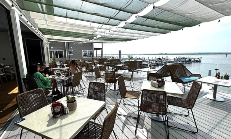
Modern Amenities and Features
Tideline Margate is designed with modern living in mind, offering a wide range of amenities that cater to diverse lifestyles. Residents can enjoy features such as swimming pools, fitness centers, landscaped gardens, and communal spaces that encourage social interaction. These amenities are thoughtfully integrated to enhance daily living and promote a sense of community.
The residences themselves are built with high-quality materials and contemporary finishes. Open floor plans, large windows, and energy-efficient designs create a comfortable and inviting atmosphere. Tideline Margate also prioritizes safety and convenience, with secure entry systems and ample parking facilities for residents and guests.
Lifestyle and Community Experience
Living in Tideline Margate means embracing a lifestyle that combines relaxation with activity. The community is designed to foster connections among residents, making it an ideal place for families, professionals, and retirees alike. Regular community events and shared spaces encourage social engagement and a sense of belonging.
The surrounding area offers plenty of opportunities for outdoor activities, from parks and walking trails to nearby beaches. Residents can enjoy a healthy and active lifestyle while taking advantage of the natural beauty that defines coastal living. Tideline Margate truly captures the essence of a balanced and fulfilling lifestyle.
Real Estate Value and Investment Potential
Tideline Margate is not only a great place to live but also a promising investment opportunity. The growing demand for properties in Margate has contributed to increasing property values, making this development an attractive option for investors. Its modern design and desirable location add to its long-term appeal.
Investors are particularly drawn to Tideline Margate because of its potential for rental income and appreciation. With more people seeking high-quality housing in well-connected areas, properties within this development are likely to maintain strong demand. This makes Tideline Margate a smart choice for those looking to invest in real estate.
Nearby Attractions and Entertainment
One of the key advantages of Tideline Margate is its proximity to a variety of attractions and entertainment options. Residents can easily access shopping malls, restaurants, and cultural venues, ensuring that there is always something to do. The nearby city of Fort Lauderdale is particularly popular for its vibrant atmosphere and scenic beaches.
In addition to urban attractions, the area also offers natural beauty and outdoor recreation opportunities. Parks, waterfront areas, and golf courses provide options for relaxation and leisure. This combination of entertainment and nature makes Tideline Margate an appealing destination for people of all ages.
Why Choose Tideline Margate?
Choosing Tideline Margate means investing in a lifestyle that prioritizes comfort, convenience, and quality. The development stands out for its thoughtful design, modern amenities, and strong sense of community. It offers a unique living experience that caters to both practical needs and personal preferences.
Moreover, the location within Broward County ensures access to essential services, employment opportunities, and recreational activities. Whether you are looking for a permanent residence or a vacation home, Tideline Margate provides an ideal setting that meets a wide range of expectations.
Conclusion
Tideline Margate represents the perfect blend of modern living and coastal charm. With its prime location in Margate, high-quality amenities, and strong investment potential, it has become a top choice for homebuyers and investors alike. The development offers a balanced lifestyle that combines relaxation, convenience, and community living.
As demand for well-designed residential spaces continues to grow, Tideline Margate stands out as a forward-thinking development that meets the needs of today’s residents. Whether you are planning to move, invest, or explore new opportunities, Tideline Margate is worth considering.
FAQs Tideline Margate
What is Tideline Margate?
A modern residential community offering coastal-style living.
Where is Tideline Margate located?
It is located in Margate, Florida.
Is Tideline Margate good for investment?
Yes, it offers strong growth and rental potential.
What amenities are available?
Pools, fitness centers, gardens, and community spaces.
Is Tideline Margate family-friendly?
Yes, it is suitable for families and professionals.
Are there nearby attractions?
Yes, beaches, parks, and entertainment options nearby.
How is accessibility in Tideline Margate?
It has excellent connectivity to major nearby cities.
What makes Tideline Margate unique?
Its blend of modern design and coastal lifestyle.
Also Read: Coexist Gaming